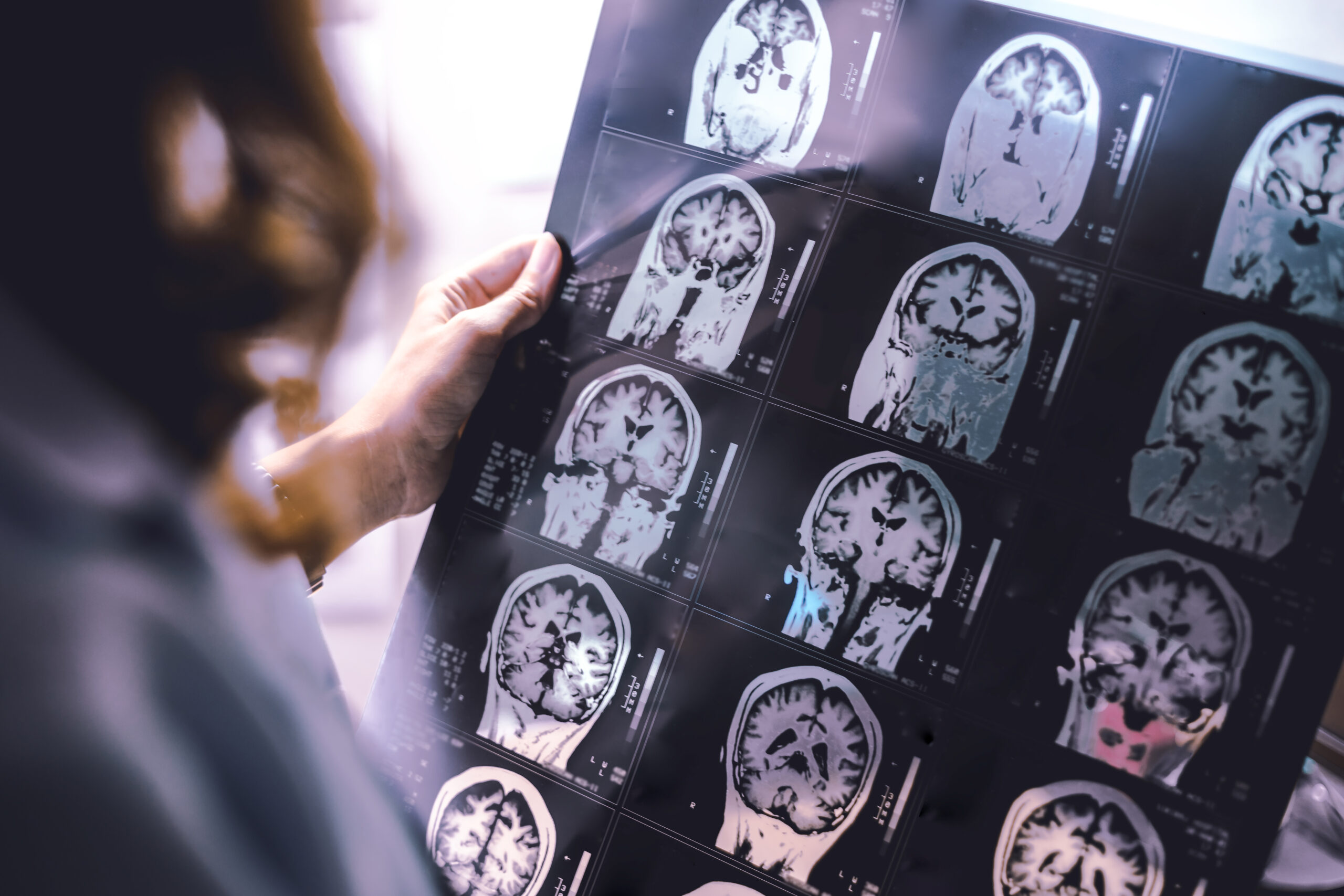
Brain and Spinal Tumours
Gliomas, meningiomas, pituitary tumours, primary CNS lymphoma and spinal tumours present in different ways but share a common need: prompt recognition, precise diagnosis and coordinated, multidisciplinary care.

Gliomas (astrocytomas, GBM, oligodendrogliomas)
What they are: Primary brain tumours arising from glial cells; behaviour ranges from slow-growing to highly aggressive.
Symptoms: New or changing headaches, seizures, progressive weakness, language or personality change, visual disturbance.
Assessment: Neurological examination and MRI with contrast. Tissue diagnosis (biopsy or resection) clarifies molecular markers (e.g., IDH status, 1p/19q codeletion, MGMT), which guide prognosis and treatment.
Treatment: Maximal safe surgical resection where feasible, followed by tailored radiotherapy and/or chemotherapy. Seizure control, steroids for swelling (short term) and structured neuro-rehabilitation are integral. Clinical trial options may be discussed

Meningiomas
What they are: Usually benign, extra-axial tumours arising from the meninges.
Symptoms: Often incidental; when symptomatic, headaches, seizures or focal deficits due to local pressure.
Assessment: MRI to define size, location and relation to venous sinuses.
Treatment: Either monitor and rescan (for small, asymptomatic lesions) or microsurgical removal/stereotactic radiosurgery when growth or symptoms warrant intervention.
Pituitary tumours
What they are: Hormone-secreting (e.g., prolactin, growth hormone, ACTH) or non-functioning lesions in the sella.
Symptoms: Headache, visual field loss (classically bitemporal), menstrual or sexual dysfunction, galactorrhoea, weight/skin changes or fatigue.
Assessment: Detailed pituitary hormone profile, MRI pituitary and formal visual field testing.
Treatment: Dopamine agonists are first-line for many prolactinomas. Others may require transsphenoidal surgery and, selectively, radiotherapy. Lifelong endocrine follow-up ensures safe hormone replacement when needed.
Primary CNS lymphoma
What it is: An aggressive brain lymphoma, more common with immunosuppression but can occur in anyone.
Symptoms: Subacute cognitive change, focal deficits, visual or speech disturbance, seizures.
Assessment: MRI, targeted bloods and biopsy. (Steroids can obscure biopsy results and are used cautiously until tissue is obtained, unless clinically essential.)
Treatment: Specialist chemotherapy-based regimens (often high-dose methotrexate-centred), with supportive care for raised pressure and seizures; radiotherapy may be used in selected scenarios.
Spinal tumours (intramedullary, extramedullary, extradural)
What they are: Lesions within or around the spinal cord that may compress neural structures.
Symptoms: Back/neck pain with bilateral limb numbness or weakness, gait imbalance, band-like torso sensation, and bladder/bowel or sexual dysfunction.
Assessment: Urgent whole-spine MRI with contrast to define the level and cause.
Treatment: Decompression and tumour management via neurosurgery and oncology as indicated; short-course steroids are used in malignant cord compression. Rehabilitation, spasticity control and falls prevention follow.
How Dr Francesco Manfredonia can help
-
Rapid triage & diagnosis: targeted examination with fast-track MRI, seizure management and safe steroid use where appropriate.
-
Joined-up pathways: close coordination with neurosurgery, neuroradiology, oncology, radiotherapy and endocrinology to deliver the right treatment at the right time.
-
Personalised support: clear explanations, written plans, driving/work guidance, and structured neuro-rehabilitation for cognition, speech and mobility.
-
Long-term follow-up: scan surveillance schedules, medication review, symptom control and supportive care for you and your family.
Urgent advice: call 999 for sudden weakness, new seizures, rapidly worsening headache with vomiting, or back pain with new bladder/bowel problems (possible cord compression).
FAQ
Are all brain tumours cancer?
No. Many (e.g., meningiomas) are benign or slow-growing. Management balances benefit and risk for your specific tumour type.
Do headaches mean I have a tumour?
Most headaches are not tumour-related. Red flags include progressive headaches, early-morning vomiting, or headaches with new neurological symptoms—these merit assessment.
Why is MRI preferred to CT?
MRI gives more detail of brain and spinal cord tissue, tumour boundaries and involvement of critical structures.
Will I need a biopsy?
Usually yes to confirm the diagnosis and molecular profile. Some pituitary micro-prolactinomas are treated medically first; your team will advise. In suspected CNS lymphoma, steroids are ideally delayed until after biopsy where clinically safe.
Can seizures after a tumour be controlled?
Often, yes. Modern anti-seizure medicines and tailored dosing are effective; you’ll also receive safety advice and, where applicable, guidance on fitness to drive.
What is stereotactic radiosurgery?
Highly focused radiation (often a single or few sessions) used for selected small tumours or residual disease, sparing surrounding brain.
Will treatment affect hormones or vision with pituitary tumours?
Possibly. That’s why endocrine testing and visual field checks are built into both diagnosis and follow-up.
Can spinal tumours be benign?
Yes—schwannomas and meningiomas are examples. Nevertheless, compression symptoms need timely evaluation.
How often will I be scanned?
Intervals depend on tumour type, treatment and stability. You’ll receive a personalised surveillance plan.
What are the side-effects of steroids?
Short-term steroids reduce swelling but can affect mood, sleep, appetite and blood sugar. Doses are kept as low and brief as possible, with a taper plan.
BOOK YOUR CONSULTATION
Book a consultation with Dr Francesco Manfredonia (Dr FM) for clear diagnosis, compassionate care and a plan built around your life and goals.
